Our cells and organs need oxygen from the air we breathe in order to survive and function. However, in certain disorders such as stroke, heart disease and cancer, tissues are often deprived of oxygen. This lack of oxygen, known as hypoxia, leads to the tissue damage and deregulation that are characteristic of these diseases. Understanding how tissues and organs respond to low oxygen is therefore an important question in biomedical research.
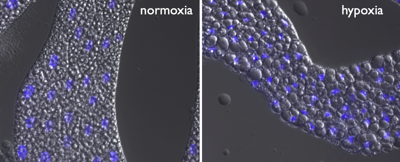
Using Drosophila as an in vivo model system, we discovered that the fly adipose tissue (the fat body) functions as a key hypoxic sensor that controls whole body hypoxia tolerance (Lee et al, 2019) . Specifically, we identified hypoxia suppression of the conserved Target-Of-Rapamycin (TOR) kinase signaling pathway as the trigger for this hypoxia response. Inhibition of TOR was required to remodel fat body lipid metabolism, which, in turn, promoted a non-autonomous, whole-body protection against the lethal effects of hypoxia. We also showed that the FOXO transcription factor is required hypoxia tolerance, in part due to its ability to induce immune-like responses via the immune transcription factor Relish/NF-KappaB (Barretto et al 2020).
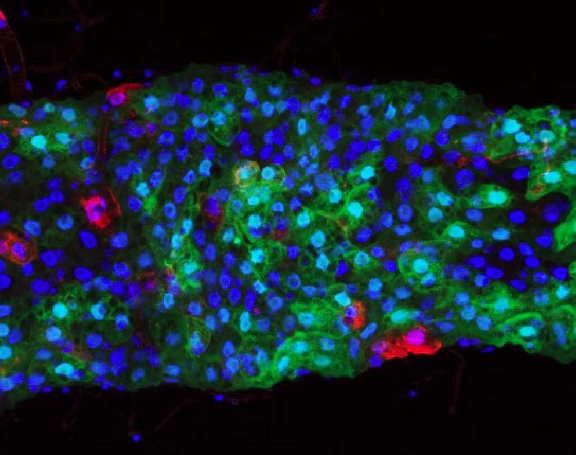
More recently, we’ve identified the gut as a second key hypoxia-sensing tissue. We found that hypoxia induces expression of the cytokine Unpaired-3 (Upd3), the fly homolog of human IL-6, in the intestine, and that gut-derived Upd3 signals to fly adpose tissue to promote hypoxia tolerance (Ding et al, 2026).
We are currently addressing several questions raised by our work:
- How do different organs sense oxygen levels and communicate with one another to produce a coordinated, whole-body response to hypoxia?
- How do changes in cell and tissue metabolism contribute to hypoxia tolerance?
- Do these hypoxic resposnses influence tissue responses in fly models of disorders associated with hypoxia such as tumorgenesis and ischemia?
\

